CARDIOVASCULAR SYSTEM [CSEC HSB & BIOLOGY]
SYLLABUS REFERENCE
CSEC Biology
- [B4.3] describe the structure and function of the circulatory system in humans;
- [B4.4] relate the structure of the components of blood to their function;
CSEC HSB
- [B3.4] relate the structures of red blood cells, phagocytes, and lymphocytes to their functions;
- [B3.5] relate the structures of the arteries, veins, and capillaries to their functions;
- [B3.6] relate the structures of the heart to their functions;
- [B3.7] explain the concept of blood pressure;
- [B3.8] describe the structure & function of the circulatory system in humans;
- [B3.11] discuss the causes & effects of heart diseases; (brief overview)
Human & other mammalian transport systems are
DOUBLE CIRCULATORY SYSTEMS.

In such a system, for every cycle, the blood passes through the heart TWICE.
The two segments of the cycle in humans is called the pulmonary & systemic circulation.

Pulmonary Circulation
Deoxygenated blood is pumped from the heart to the lungs, through the pulmonary arteries.
Oxygenated blood flows from the lungs, back to the heart in the pulmonary veins.
Systemic Circulation
Oxygenated blood is pumped from the heart to the rest of the body, via the aorta and other arteries.
Deoxygenated blood returns from the rest of the body to the heart via the vena cavae.
COMPONENTS OF THE CIRCULATORY SYSTEM

THE BLOOD VESSELS
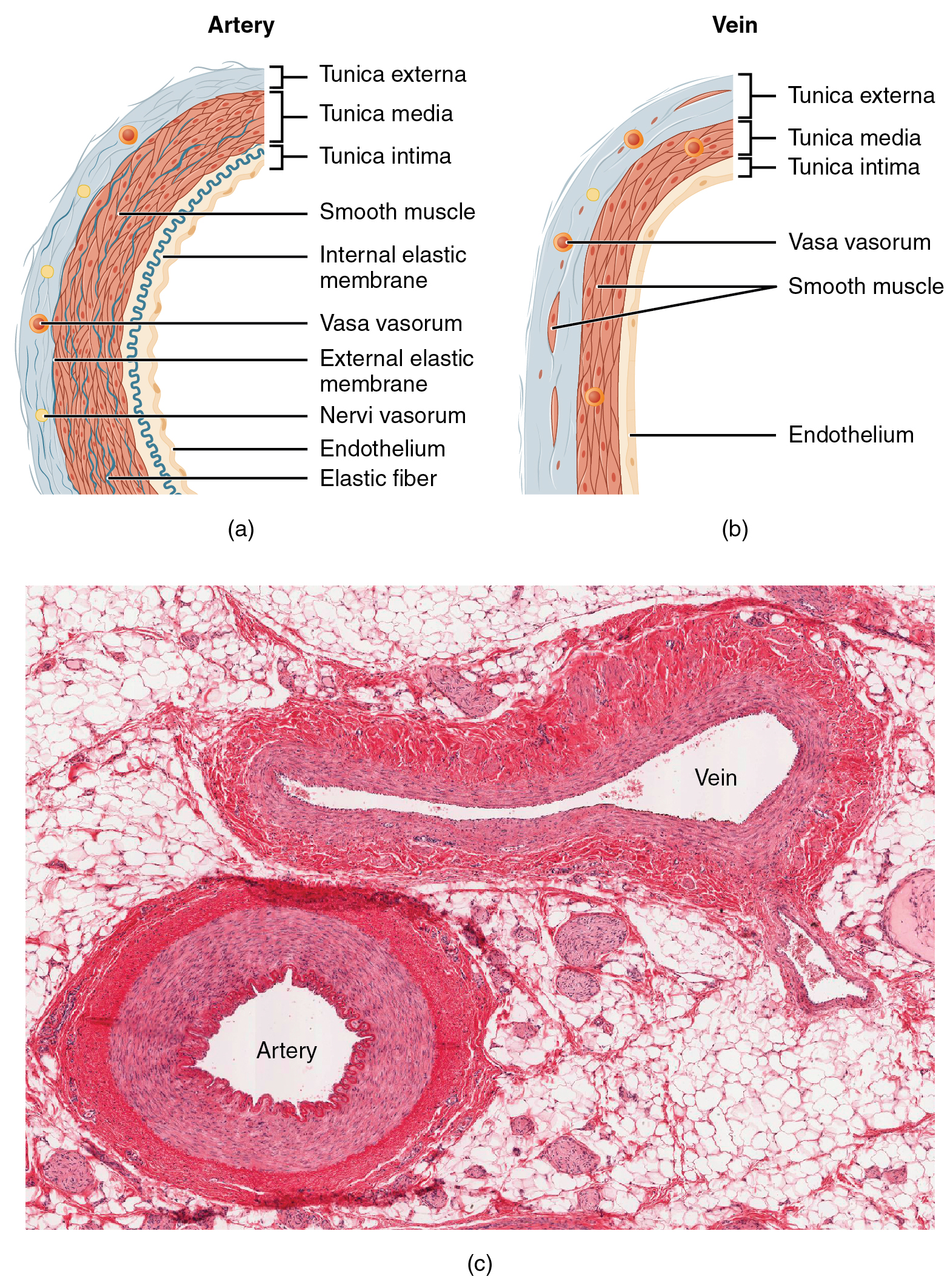
Artery
- Blood flows AWAY from the heart at high pressure.
- Elastic tissue in the walls stretches and recoils to maintain the blood pressure.
- Blood is delivered to the organs at a pressure slightly less than it left the heart.
- Walls have thick layers of muscle and fibrous tissue to withstand the high blood pressure.
Vein
- Blood flows TOWARDS the heart at low pressure.
- Vessels expand to take increasing volumes of blood, such as during exercise.
- As blood pressure is low, backflow of blood is prevented by semi-lunar valves at intervals along the veins.
- Walls have thin layers at muscle and fibrous tissue as they do not have to withstand high blood pressures.
Capillary
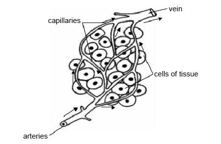
- Blood flows between smaller arteries (arterioles) and smaller veins (venules) at low pressure.
- Oxygen and carbon dioxide diffuse through the walls.
- Water and solutes, such as glucose and amino acids, pass through the walls into tissue fluid surrounding the cells.
- Walls are thin so there is a short diffusion distance between the blood and the tissue fluid.
THE HEART


THE CARDIAC CYCLE


COMPONENTS OF BLOOD

The blood consists of a pale-yellow liquid called plasma in which blood cells and platelets are suspended as they move around the body.

Plasma
Plasma is about 90% water.
It transports dissolved substances such as digested food molecules, hormones, vitamins, and mineral salts around the body.
Red Blood Cells - Erythrocytes
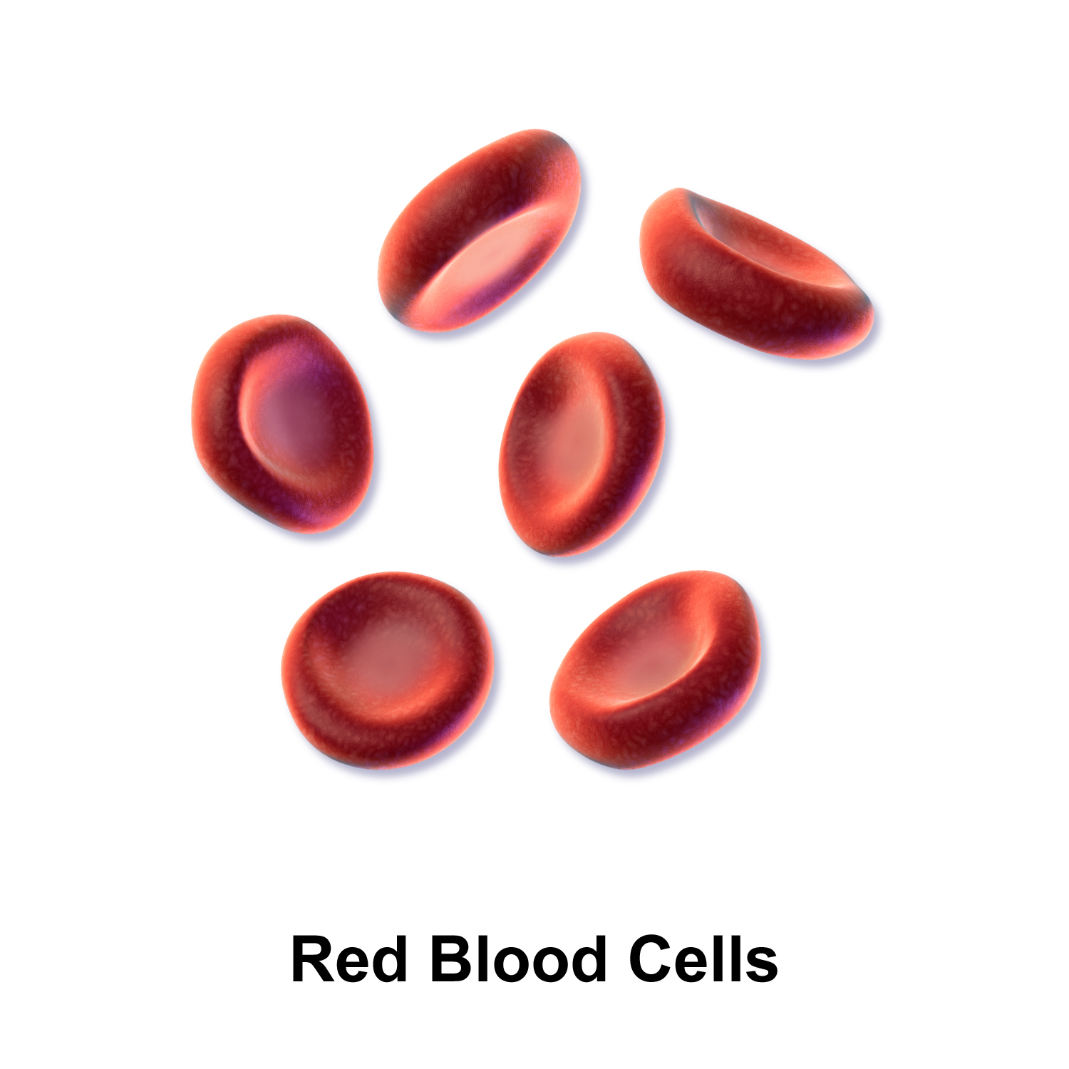
These cells are red because they contain a red pigment called haemoglobin. This pigment combines with oxygen and transports it to cells in the body.
They have a biconcave disc shape. Mature red blood cells do not have a nucleus.
They are produced in large quantities in the bone marrow at the end of the large limb bones, such as the femur.
They live for about 120 days, after which they are destroyed or broken down in the liver.
White Blood Cells - Leukocytes
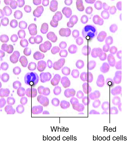
These cells are larger than red blood cells and have a nucleus.
Depending on the type of white blood cell, the nucleus is either lobed or spherical.
They do not contain haemoglobin. There is about 1 white blood cell for every 300 red blood cells.
Their main function is to defend the body against invading organisms and their products.
There are two main categories of white blood cells:
- Phagocytes
- Lymphocytes

Phagocytes
These white blood cells are able to change their shape, like an amoeba. This enables them to squeeze through the tiny gaps in the walls of capillaries.
The phagocytes destroy invading organisms, such as bacteria, by flowing around them and engulfing them. They then digest the organisms inside them.
Lymphocytes
Lymphocytes will find antigens (usually a protein) in the body that they do not recognise, such as from an invading organism. They then release chemicals called antibodies that match the shape of the antigen on the invading organism.
The antibodies will destroy the invading cells by causing them to either stick together or burst, making it easier for the phagocytes to clear them up.
Lymphocytes are also able to convert toxins (poisons), from invading organisms into less toxic substances.
Platelets
These are fragments of cells formed in the bone marrow.
Their main function is to help in the clotting of blood in open wounds.

After a blood vessel is cut, and there is some bleeding for a while, platelets near the wound cause the blood to produce thread-like proteins called fibrin. These form a mesh of fibres that traps the blood cells and eventually forms a blood clot.

This seals the wound and prevents further bleeding.
CARDIOVASCULAR DISEASES
These are the most common conditions
- Hypertension
- Heart attack
- Stroke
Hypertension (High Blood Pressure)
Hypertension occurs when the pressure of the blood passing through the blood vessels is higher than the norm.
When someone suffers from hypertension, it means that great force is exerted on the arteries, especially when the heart contracts. So their systolic reading will be much higher than 120.
Difference between the systolic reading and the diastolic reading of a blood pressure measurement.
The systolic pressure is the higher number and shows the blood pressure as the heart is contracting.
The diastolic pressure is the lower number and shows the blood pressure when the heart is relaxing.

Hypertension are caused by several factors. Two of the most prominent factors - which come from components of the circulatory system itself - are arteriosclerosis and atherosclerosis.
Arteriosclerosis
It involves the hardening of the arteries, causing them to lose elasticity.
This makes it impossible for the artery to stretch to accommodate the blood which is being forced through it.
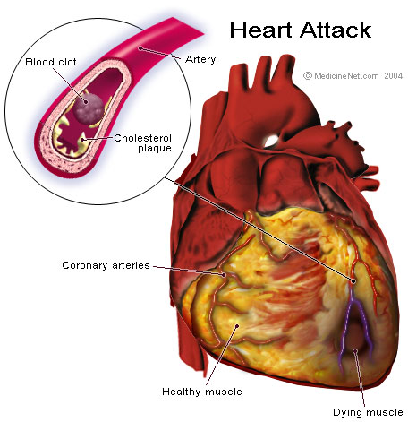
Atherosclerosis
This happens if the diameter of the artery becomes narrower due to the buildup of fatty deposits of cholesterol on the wall.
Hypertension puts greater pressure on the heart since the blood has to be pushed through a much smaller diameter.

Angina
The heart keeps pumping because it gets a continuous supply of food and oxygen from the blood passing through the coronary arteries.
If the coronary arteries harden, this will restrict the flow of blood to the heart muscles. The result is severe chest pains called angina.
Thrombosis
Sometimes the cholesterol build-up in an artery creates a very rough surface through which the blood passes. This can result in the formation of a blood clot which is released into the blood flow.
If the blood clot is large enough, it can block the narrower passages in an artery and stop the blood flow through it.
This is a condition called thrombosis.

Heart Attack
If the thrombosis occurs in the coronary artery (coronary thrombosis) this can stop the heart beating and the person has a heart attack.
To start the heart beating again, it needs to be massaged or given electrical shocks.
In the longer term, it can be treated by giving the patient a coronary bypass. This is an operation in which a piece of blood vessel is taken from the person's leg and used to create a new passage for the flow of blood to the heart muscles, bypassing the blocked area.
Stroke
A stroke occurs when there is an obstruction in the flow of blood to the brain. This deprives the brain of oxygen and nutrients, causing brain cells to die.
Strokes may result in sudden weakness of the limbs, which impedes mobility. There may also be a loss of sensation, accompanied by the inability to speak or see clearly.
There are two main types of strokes:
- Ischaemic
- Haemorrhagic

An ischaemic stroke happens when there is a blockage of the blood vessels leading to the brain, which may be caused by a blood clot.
A haemorrhagic stroke occurs when the blood vessels in the brain rupture or leak.
Note that if the symptoms of a stroke are short-lived (a few hours) then the person is said to have had a transient ischaemic attack (TIA), also called a mini-stroke.
The following persons are at high risk of having a stroke. Those diagnosed with:
- hypertension
- high cholesterol
- diabetes
- obstructive sleep apnoea