EYE STRUCTURE & FUNCTION [CSEC HSB]
SYLLABUS REFERENCE
CSEC HSB
- [B6.9] relate the internal structures of the eye to their functions;
- [B6.10] explain how images are formed in the eye;
- [B6.11] explain accommodation in the eye;
- [B6.12] discuss the causes of, and corrective measures for eye defects and diseases.
CSEC Biology
- [B7.9] relate the structure of the human eye to its functions as a sense organ;
- [B7.10] explain accommodation; sight defects and the corrections of each;
The Eye is a Sense Organ That Contains Light Receptors
EXTERNAL STRUCTURE OF THE EYE

Eyelid
- Protects the eye
- Blinking keeps surface of eye and conjunctiva moist
Conjunctiva
- Thin, transparent epithelium covering the eyeball
- Secretes mucus to lubricate, clean and maintain moisture
Iris
- Pigmented. This is where eye colour is located.
- Has radial and circular muscles which regulate the amount of light entering the eye through the pupil.
Pupil
Round hole at the centre of the iris, through which light enters
Sclera
- Tough, opaque, non-elastic layer on the outside of the eyeball.
- Helps to maintain shape of eyeball.
INTERNAL STRUCTURE OF THE EYE

Cornea
Transparent, curved layer bends light to converge at lens.
Lens
- Soft, transparent and elastic
- Focuses light on the retina
Ciliary body/muscle
Muscle which help the lens alter its shape after focusing
Aqueous Humour
- Transparent water liquid in front of the iris and pupil
- Medium for diffusion of oxygen and nutrients
- Refracts light and maintains shape of eyeball
Vitreous Humour
- Transparent jelly, in the space between lens and retina
- Refracts light and maintains shape of eyeball
Retina
- Light-sensitive layer of the eye
- Has photoreceptor cells (rods and cones)
Yellow Spot / Fovea Centralis
High cone density for night vision
Blind Spot
- Where nerve fibres connecting rods and cones leave the eye
- Has no photoreceptor cells
Choroid
- Middle layer with black pigments
- Prevents internal reflection of light
Optic Nerve
- Made of sensory nerve fibres
- Transmit information from eye to brain
HOW DOES THE EYE WORK?
Accommodation
- Images are focused onto the retina by the adjustments made to the eye.
- Ciliary muscles contract & relax to focus on near and distant objects.
- This contraction & relaxation changes the lens curvature.
- Change in curvature in turn changes the distance of focus (focal length) of the lens.

Focusing on Near Objects
- Ciliary muscles contract
- Tension in suspensory ligament reduces
- Lens becomes more convex
- Pupil size reduced
Focusing on Distant Objects
- Ciliary muscles relax
- Tension in suspensory ligaments increases
- Lens become more concave
- Pupil size increased
Response to Light

- The pupil controls the amount of light entering the eye
- Light enters as a stimulus to regulate the size of the pupil
- Both eyes adjust the same way to the light conditions.
EYE DISORDERS

Myopia (nearsightedness) and hypertropia (farsightedness) can be fixed using lenses, placed in eye glasses.
Nearsightedness and its Correction

Farsightedness and its Correction

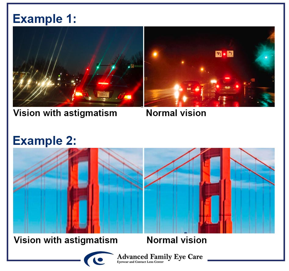
Astigmatism
This is an imperfection in the curvature of your eye's cornea or lens.
With astigmatism, the eyeball has an egg or oval shape, instead of a rounded shape. As a result, vision for BOTH near and far objects is blurry and distorted.
.png/290px-Astigmatism_(Eye).png)
Glaucoma

This is a disease that damages the eye's optic nerve. It usually happens when fluid builds up in the front part of the eye. That extra fluid increases the pressure in one's eye, damaging the optic nerve.
Glaucoma is a leading cause of blindness for people over 60 years old. But blindness from glaucoma can often be prevented with early treatment.

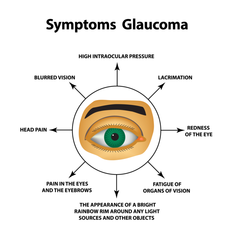
Glaucoma is usually controlled with eyedrop medicine.
Used every day, these eye drops lower eye pressure.
Some do this by reducing the amount of aqueous fluid the eye makes.
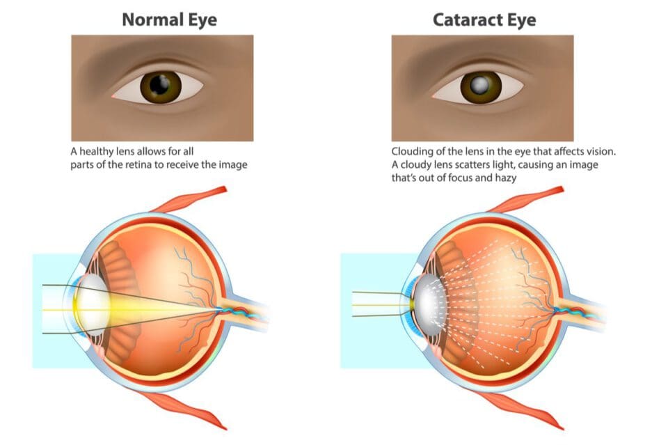
Cataract
A cataract is a clouding of the normally clear lens of the eye.

For people who have cataracts, seeing through cloudy lenses is a bit like looking through a frosty or fogged-up window.
Most cataracts develop when aging or injury changes the tissue that makes up the eye's lens.
Proteins and fibers in the lens begin to break down, causing vision to become hazy or cloudy.
At first, stronger lighting and eyeglasses can help a patient deal with cataracts. But if impaired vision interferes with one's usual activities, one might need cataract surgery. Fortunately, cataract surgery is generally a safe, effective procedure.
How Diabetes Affects the Eyes
Both Type 1 and Type 2 diabetes causes high blood sugar concentrations.
High concentrations of sugar in the blood damages the arteries that carry oxygenated blood from the heart to the various organs & tissues in the body. This includes the tiny blood vessels (arterioles and capillaries) that provide nutrients and oxygen to the eye, and especially the retina. Also, parts of the eye itself is exposed to high concentration of sugar.
The result is a range of diabetic eye issues, which include the following:
- Blurred vision
- Diabetic retinopathy
- Glaucoma
- Macular oedema
- Cataracts
Blurred Vision
This is usually one of the first diabetic eye symptoms and issues.
Exposure to high sugar concentrations causes swelling of the lens. This, in turn, produces blurry vision.
The good news is that this condition can be reversed, simply by controlling blood sugar levels. Once blood sugar levels are successfully controlled, it can take up to six weeks for the lens (and one's vision) to return to normal.
If the blurry vision is intense enough to interfere with a person's ability to function, then one may need a change to eyeglass prescription. This would be a temporary change until vision returns to normal.
Diabetic Retinopathy
This is a diabetic eye disease.
This occurs when blood vessels in the retina swell, leak fluid or bleed due to damage caused by high blood sugar concentrations. Other abnormalities include growth of extra blood vessels in the eye, and closing off of existing blood vessels to the retina.
Early states may be symptomless. However, as the disease progresses one can start to experience the following symptoms:
- Blurred vision
- Vision that becomes blurry, then suddenly returns to clear
- Sudden appearance of or considerable increase in eye floaters
- Poor night vision
- Blank spots or dark shadows in one's field of vision
If left untreated, this disease can eventually lead to permanent blindness.
Macular Oedema
This can be a consequence of diabetes and diabetic retinopathy.
This is the collection of fluid - from leaking blood vessels - in the macula (aka yellow body). This is the part of the retina to produces the most detailed vision.
Initial symptoms is painless blurry vision. It can lead to permanent loss of visual acuity (aka visual sharpness).